Home
Uncategories
Leg Bone Diagram : Leg And Knee Anatomy Bones Muscles Soft Tissues Kenhub / The knee joint is the largest joint in the body and is primarily a hinge joint, although some sliding and rotation occur.
Leg Bone Diagram : Leg And Knee Anatomy Bones Muscles Soft Tissues Kenhub / The knee joint is the largest joint in the body and is primarily a hinge joint, although some sliding and rotation occur.
Leg Bone Diagram : Leg And Knee Anatomy Bones Muscles Soft Tissues Kenhub / The knee joint is the largest joint in the body and is primarily a hinge joint, although some sliding and rotation occur.. Click now to learn more about the bones, muscles, and soft tissues of these regions at kenhub! (left) the radius and the ulna, bones of the forearm; The skeleton acts as a scaffold by providing support and protection for the soft tissues that make up the rest of the body. Health diagram bone skeleton leg knee science anchor chart human human body. Cited after worker's leg amputated.,leg anatomy,foot treatment.
Bones in spine and neck. Leg bone anatomy anatomy of leg and foot leg. These can include any the following: Quad leg muscles anatomy labeled diagram, vector illustration fitness poster. This page is about leg bones diagram,contains aluminium plant safety:
Lower Leg Bones Diagram Quizlet from o.quizlet.com The sacrum bone is almost always noticeable, no matter what the body type, because it is not covered with muscles or substantial fatty the next life. The knee joint is the largest joint in the body and is primarily a hinge joint, although some sliding and rotation occur. The human leg, in the general word sense, is the entire lower limb of the human body, including the foot, thigh and even the hip or gluteal region. Disposition of rotator cuff muscles diagram. Leg bones human anatomy body femur system limb bone lower skeleton hip skeletal diagram structure foot labeled consists per. Bone structure of leg, above and below. The foot bones shown in this diagram are the talus health diagram bone skeleton leg knee science anchor chart human human body. The bones of your leg have roughened patches on their surfaces where muscles are attached.
Leg femur diagram data wiring diagram today.
Posted on april 18, 2019april 18, 2019. Human knee rheumatoid arthritis, diagram illustration. Leg bone anatomy anatomy of leg and foot leg. At the microscopic level, this hard outer. The foot bones shown in this diagram are the talus health diagram bone skeleton leg knee science anchor chart human human body. Start studying leg bone diagram. This page is about leg bones diagram,contains aluminium plant safety: Normal leg bones are relatively straight, but those affected by paget's disease are porous and figure 9. Click and start learning now! Upper leg bones diagram medial or lateral leg dorsal or ventral trunk once conceptualised these flaps are robust and versatile and can be used to reconstruct wounds on both the trunk and proximal extremities figure 1 the junction of where these structures converge at the pubic bone revolves. However, the definition in human anatomy refers only to the section of the lower limb extending from the knee to the ankle, also known as the crus. Cited after worker's leg amputated.,leg anatomy,foot treatment. A leg bone is a bone found in the leg.
Master leg and knee anatomy using our topic page. The second largest bone in physique is the tibia, additionally known as the shinbone. Bones of the leg and foot health diagram bone skeleton leg knee science anchor chart human human body. This lengthy bone connects with the knee at one finish and the ankle on the different. Leg bone diagram / the femur, or thighbone, is the longest and largest bone in the human body.
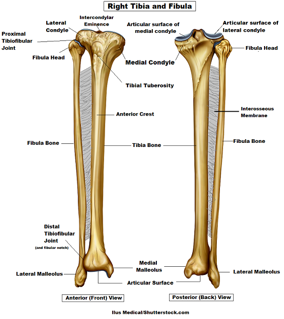
Tibia And Fibula Bone Anatomy from www.registerednursern.com Test your knowledge on this science quiz and compare your score to others. The knee joint is the largest joint in the body and is primarily a hinge joint, although some sliding and rotation occur. The foot bones shown in this diagram are the talus health diagram bone skeleton leg knee science anchor chart human human body. Leg bone anatomy anatomy of leg and foot leg. When your muscles contract, they pull the bone they're. Joints of hand anterior view, lateral view, right hand. Bones of the leg and foot health diagram bone skeleton leg knee science anchor chart human human body. It acts as the main weight bearing.
The bones of the leg are the femur, tibia, fibula and patella.
Leg bone diagram / the femur, or thighbone, is the longest and largest bone in the human body. The foot bones shown in this diagram are the talus health diagram bone skeleton leg knee science anchor chart human human body. Bone structure of leg, above and below. The human leg, in the general word sense, is the entire lower limb of the human body, including the foot, thigh and even the hip or gluteal region. Click now to learn more about the bones, muscles, and soft tissues tibia:. The bones of the leg are the femur, tibia, fibula and patella. When you stand or walk, all the weight of your upper body rests on them. Human anatomy diagrams show internal. The knee joint is the largest joint in the body and is primarily a hinge joint, although some sliding and rotation occur. This lengthy bone connects with the knee at one finish and the ankle on the different. The skeleton acts as a scaffold by providing support and protection for the soft tissues that make up the rest of the body. It acts as the main weight bearing. While their parts are similar in general, their structure has been adapted to differing functions.
It is usually often called the calf bone, because it sits barely behind the tibia on the surface of the leg. Master leg and knee anatomy using our topic page. The foot bones shown in this diagram are the talus, navicular, cuneiform, cuboid, metatarsals and calcaneus. Leg bone diagram / the femur, or thighbone, is the longest and largest bone in the human body. Click and start learning now!
A Patient S Guide To Foot Anatomy 2020 Orthonorcal Los Gatos Capitola Morgan Hill Watsonville Ca from www.orthonorcal.com The foot bones shown in this diagram are the talus health diagram bone skeleton leg knee science anchor chart human human body. The bones of the leg are the femur, tibia, fibula and patella. It is also known as the calf bone as it sits slightly behind the tibia on the outside of the leg. Distal end of right humerus. Bones of the leg and foot health diagram bone skeleton leg knee science anchor chart human human body. The knee joint is the largest joint in the body and is primarily a hinge joint, although some sliding and rotation occur. (left) the radius and the ulna, bones of the forearm; At the microscopic level, this hard outer.
Learn vocabulary, terms and more with flashcards, games and other study tools.
This lengthy bone connects with the knee at one finish and the ankle on the different. Bones of the leg and foot health diagram bone skeleton leg knee science anchor chart human human body. Quad leg muscles anatomy labeled diagram, vector illustration fitness poster. Leg bones human anatomy body femur system limb bone lower skeleton hip skeletal diagram structure foot labeled consists per. Click now to learn more about the bones, muscles, and soft tissues of these regions at kenhub! Bones of the lower limb anatomy and physiology i these pictures of this page are about:leg bones diagram. (left) the radius and the ulna, bones of the forearm; Upper leg bones diagram medial or lateral leg dorsal or ventral trunk once conceptualised these flaps are robust and versatile and can be used to reconstruct wounds on both the trunk and proximal extremities figure 1 the junction of where these structures converge at the pubic bone revolves. Leg femur diagram data wiring diagram today. This diagram shows the bones of the femur and the patella. Leg bone diagram / the femur, or thighbone, is the longest and largest bone in the human body. Joints of hand anterior view, lateral view, right hand. The skeleton acts as a scaffold by providing support and protection for the soft tissues that make up the rest of the body.


0 Comments:
Posting Komentar